Simple Compact Bone Diagram : The Anatomy and Physiology of Animals/Skeleton Worksheet ... : The remainder is cancellous bone, which has a spongelike appearance with numerous large spaces and is found in the.
byAdmin-
0
Simple Compact Bone Diagram : The Anatomy and Physiology of Animals/Skeleton Worksheet ... : The remainder is cancellous bone, which has a spongelike appearance with numerous large spaces and is found in the.. Compact bone is the denser, stronger of the two types of bone tissue (). Structure of human bones explained. Under the periosteum is a thin layer of compact bone (often called cortical bone). Provides support/protection for the body. (b) in this micrograph of the osteon, you can clearly see the concentric lamellae and central canals.
Skeletal diagram of foot a bone inside marrow stem cell is type. Compact bone, also called cortical bone, dense bone in which the bony matrix is solidly filled with organic ground substance and inorganic salts, leaving only tiny spaces (lacunae) that contain the osteocytes, or bone cells.compact bone makes up 80 percent of the human skeleton; Compare and contrast the following terms a) epiphysis vs. Each osteon is composed of concentric rings of calcified matrix. This video describes the microscopic anatomy of compact bone.
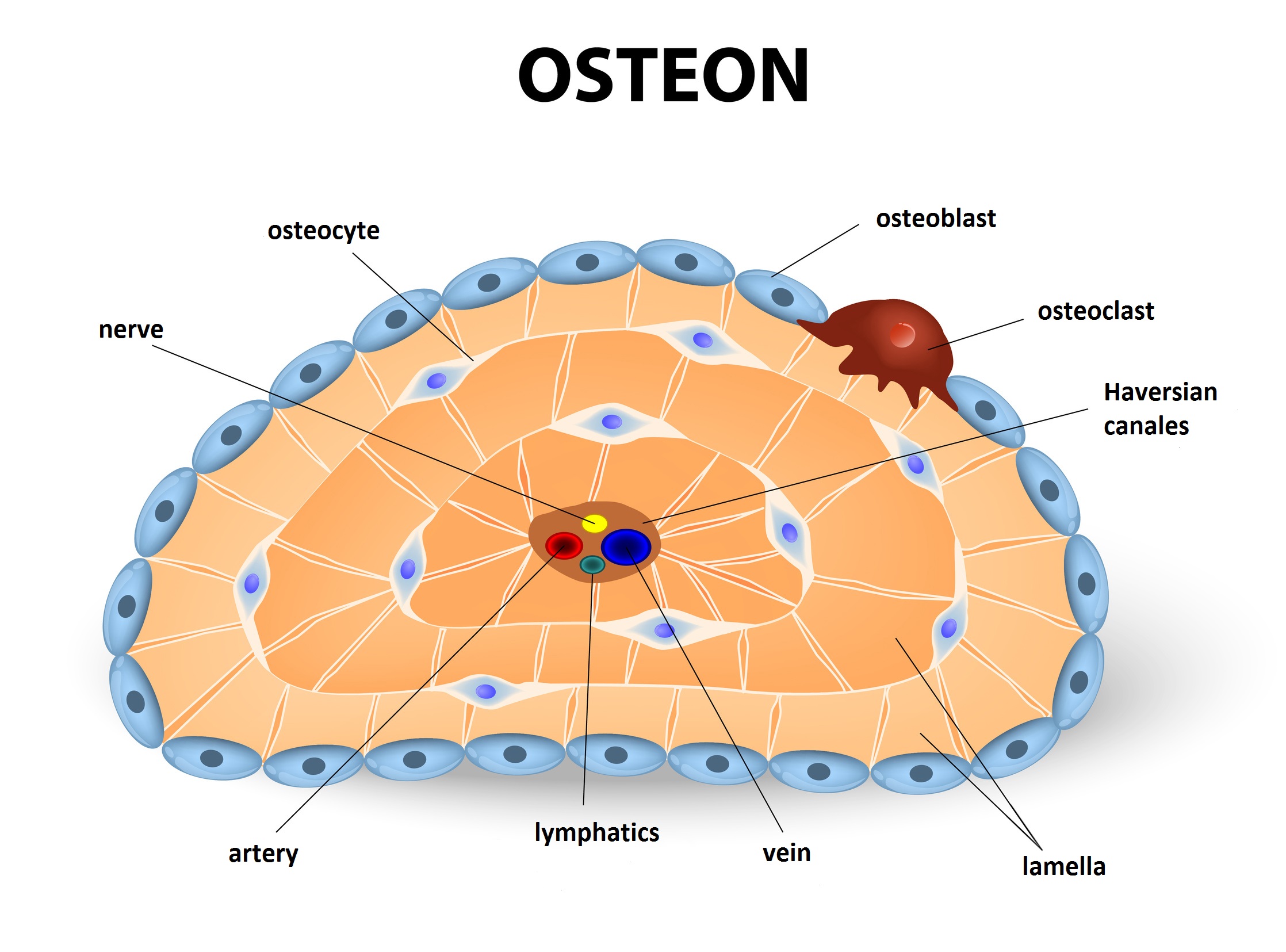
Bones: Fundamentals of anatomy for physicians | Lecturio from blog.lecturio.com Many tiny cells called osteocytes live in small spaces in the matrix and help to maintain the strength and integrity of the compact bone. There is a printable worksheet available for download here so you can take the quiz with pen and paper. The diagram above shows a longitudinal view of an osteon. It coats the inner compact bone and the trabeculae of the spongy bone. (b) in this micrograph of the osteon, you can clearly see the concentric lamellae and central canals. The foot bones shown in this diagram are the talus, navicular, cuneiform, cuboid, metatarsals and calcaneus. (b) in this micrograph of the osteon, you can clearly see the concentric lamellae and central canals. Cells human bones names wiring diagram database.
Compact bone is made of a matrix of hard mineral salts reinforced with tough collagen fibers.
This video describes the microscopic anatomy of compact bone. As seen in the image below, compact bone forms the cortex, or hard outer shell of most bones in the body. (b) in this micrograph of the osteon, you can clearly see the concentric lamellae and central canals. The microscopic structural unit of compact bone is called an osteon, or haversian system. This provides the bones strength and consists of tightly stacked layers of bone which appear to form a solid section. Diaphysis b) spongy bone vs. Bone is made up of two base componets: Each osteon is composed of concentric rings of calcified matrix. It can be found under the periosteum and in the diaphyses of long bones, where it provides support and protection. Compact bone, also called cortical bone, dense bone in which the bony matrix is solidly filled with organic ground substance and inorganic salts, leaving only tiny spaces (lacunae) that contain the osteocytes, or bone cells.compact bone makes up 80 percent of the human skeleton; The foot bones shown in this diagram are the talus, navicular, cuneiform, cuboid, metatarsals and calcaneus. Learn vocabulary, terms and more with flashcards, games and other study tools. Structure of human bones explained.
Compact bone, as opposed to spongy bone, is made of cylindrical units, called osteons, that are tightly formed together. Bone is made up of two base componets: Compact bone, dense bone in which the bony matrix is solidly filled with organic ground substance and inorganic salts, leaving simple compact bone diagram labeled : Compact bone, also called cortical bone, dense bone in which the bony matrix is solidly filled with organic ground substance and inorganic salts, leaving only tiny spaces (lacunae) that contain the osteocytes, or bone cells.compact bone makes up 80 percent of the human skeleton; Compact bone is made of a matrix of hard mineral salts reinforced with tough collagen fibers.
Structure and Function of the Haversian System Explained ... from www.buzzle.com Learn vocabulary, terms and more with flashcards, games and other study tools. The foot bones shown in this diagram are the talus, navicular, cuneiform, cuboid, metatarsals and calcaneus. White tissue composed of osteocytes in a matrix of calcium salts. Cells human bones names wiring diagram database. Start studying leg bone anatomy. It makes up the outer cortex of all bones and is in immediate contact with the periosteum. The compact bones form the hard exterior of the bones, whereas the spongy bones have several pores that are filled with nerves and blood vessels. (b) in this micrograph of the osteon, you can clearly see the concentric lamellae and central canals.
As seen in the image below, compact bone forms the cortex, or hard outer shell of most bones in the body.
(b) in this micrograph of the osteon, you can clearly see the concentric lamellae and central canals. Under the periosteum is a thin layer of compact bone (often called cortical bone). Terms in this set (8) spongy bone (contains red marrow) compact bone (has osteons) osteon. As seen in the image below, compact bone forms the cortex, or hard outer shell of most bones in the body. However, they do contain osteons, which are like canals, providing passageways through the hard bone matrix. This video describes the microscopic anatomy of compact bone. The foot bones shown in this diagram are the talus, navicular, cuneiform, cuboid, metatarsals and calcaneus. The remainder is cancellous bone, which has a spongelike appearance with numerous large spaces and is found in the. The fishbone template is a simple visualization of a problem's causes and as the name implies, the diagram has the appearance of a fish skeleton, where each bone represents a category of a root cause. Bone marrow diagram, compact bone diagram quiz, compact bone slide labeled, diagram long bone, labeled compact bone model, human anatomy, bone marrow diagram, compact bone diagram quiz, compact bone slide labeled, diagram long bone, labeled compact bone model. Definition and functions the endosteum is a structure in the middle of bone tissue and bone marrow. The compact bones form the hard exterior of the bones, whereas the spongy bones have several pores that are filled with nerves and blood vessels. White tissue composed of osteocytes in a matrix of calcium salts.
It is also called osseous tissue or cortical bone and it provides structure and support for an organism as part of its skeleton, in addition to being a location for the storage of minerals like calcium.about 80% of the weight of the human skeleton comes from. Compact bone, dense bone in which the bony matrix is solidly filled with organic ground substance and inorganic salts, leaving simple compact bone diagram labeled : Compare and contrast the following terms a) epiphysis vs. There is a printable worksheet available for download here so you can take the quiz with pen and paper. Compact bone is the denser, stronger of the two types of osseous tissue (figure 6.3.6).
human arm bone microstructure - Google Search | Human ... from i.pinimg.com Compact bone is the denser, stronger of the two types of osseous tissue (figure 6.3.6). A) spongy bone b) diaphysis c) yellow bone marrow d) epiphysis e) red bone marrow f) medullary cavity g) periosteum h) articular cartilage 3. (b) in this micrograph of the osteon, you can clearly see the concentric lamellae and central canals. The labels include periosteum, compact bone, nutrient artery & vein, medullary cavity, yellow bone marrow, endosteum, epiphyseal line, and spongy bone with red bone marrow. Diaphysis b) spongy bone vs. Human skeleton long bones of arms and legs britannica. (b) in this micrograph of the osteon, you can clearly see the concentric lamellae and central canals. (b) in this micrograph of the osteon, you can clearly see the concentric lamellae and central canals.
(b) in this micrograph of the osteon, you can clearly see the concentric lamellae and central canals.
Learn vocabulary, terms and more with flashcards, games and other study tools. It is made up … Compact bone, dense bone in which the bony matrix is solidly filled with organic ground substance and inorganic salts, leaving simple compact bone diagram labeled : This video describes the microscopic anatomy of compact bone. The labels include periosteum, compact bone, nutrient artery & vein, medullary cavity, yellow bone marrow, endosteum, epiphyseal line, and spongy bone with red bone marrow. The compact bones form the hard exterior of the bones, whereas the spongy bones have several pores that are filled with nerves and blood vessels. (b) in this micrograph of the osteon, you can clearly see the concentric lamellae and central canals. Bone is made up of two base componets: The remainder of the bone is formed by cancellous or spongy bone. (b) in this micrograph of the osteon, you can clearly see the concentric lamellae and central canals. (b) in this micrograph of the osteon, you can clearly see the concentric lamellae and central canals. Cells human bones names wiring diagram database. 13 photos of the compact bone diagram labeled.
Terms in this set (8) spongy bone (contains red marrow) compact bone (has osteons) osteon compact bone diagram. However, they do contain osteons, which are like canals, providing passageways through the hard bone matrix.